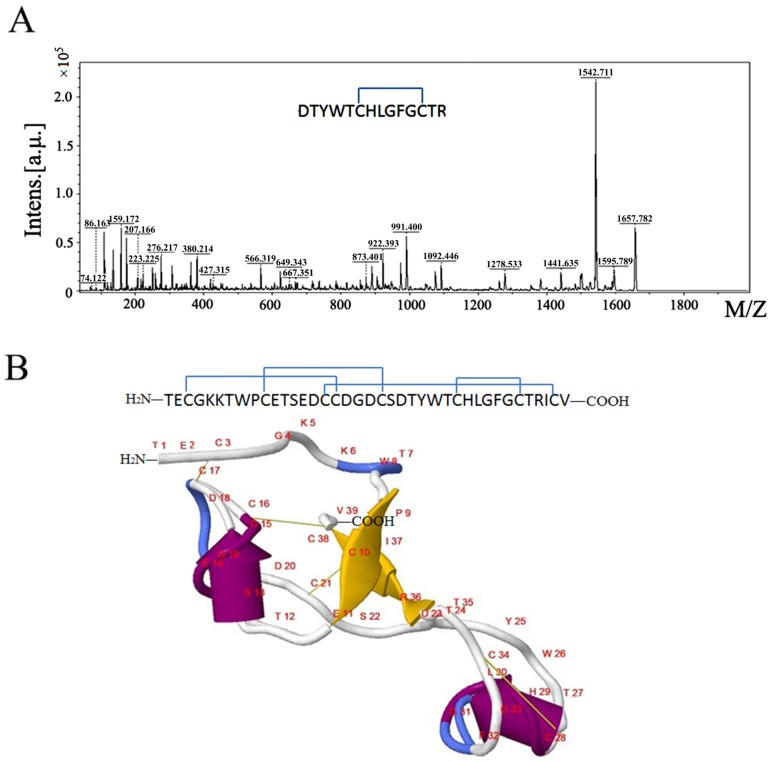
Figure 3.
The MS spectra for mapping of the disulfide bond between Cys28 and Cys34 and the predicted secondary structure of µ-TRTX-Hl1a. (A) The MS/MS spectrum of peptide DTYWTCHLGFGCTR; and (B) the secondary structure and disulfide bonds of µ-TRTX-Hl1a predicted by Phyre 2. In the figure, the disulfide bonds are indicated in yellow line, alpha helices were shown as “yellow rockets”, beta strands were shown as “purple planks”; arrowheads point towards the carboxyl termini, random coils were colored in white, and turns were colored in blue.

