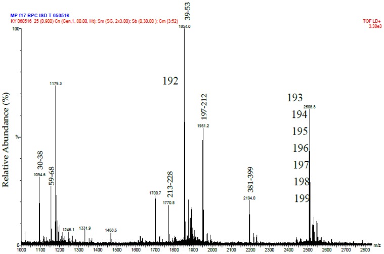
Figure 5.
MALDI-MS spectrum of tryptic peptides from purified Daborhagin. The data was obtained by digesting the R2 fraction from RP-HPLC with trypsin. The numbers above the peptides masses indicates the residue numbers for the peptides matched to the sequence of Daborhagin K (VM3DK_DABRR) (B8K1W0). All m/z values are for the M + H+ ions. The ions at 1854 and greater have been labeled with the m/z value for the ion containing one carbon as the C13 isotope.

