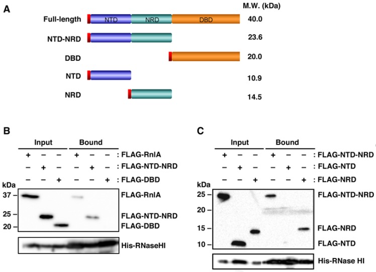
Figure 1.
Interaction of RNase HI with domains of RnlA. (A) NTD, NRD, and DBD, three domains of RnlA, are shown. The N-terminal red box represents the FLAG-tag used for detection, and the molecular weights of full-length RnlA and its derivatives are indicated on the right; (B) ΔrnlAB ΔrnhA cells harboring pQE80L-rnhA plus one of pBAD33-Flag-rnlA, pBAD33-Flag-NTD-NRD, and pBAD33-Flag-DBD; or (C) ΔrnlAB ΔrnhA cells harboring pQE80L-rnhA plus one of pBAD33-Flag-NTD-NRD, pBAD33-Flag-NTD, and pBAD33-Flag-NRD, were grown in LB medium at 30 °C until the OD600 reached 0.5, and treated with 0.06 mM IPTG and 0.05% arabinose for 45 min to induce His-tagged RNase HI and FLAG-tagged proteins. Cell extracts were subjected to pull-down with Ni-NTA beads. Input and bound fractions were analyzed by western blot with antibodies against FLAG-tag (upper panel) and His-tag (lower panel). Duplicate experiments in figure (B) and triplicate experiments in figure (C) were performed and similar results were obtained for each experiment. A representative result is shown as each figure.

