Figure 2.
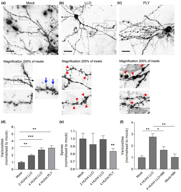
Neurite morphology in acute mouse brain slices after LLO challenge: (a) neurons in acute brain slices (PD 10–14) stained with DiI, visualizing the whole neurite tree (dendrites and axons) of intact neurons. In mock-treated samples, normal configuration of dendrites with only accidental widening in the form of varicosity (red arrow) is observed; (b) a neuron in the LLO-treated slice (4 HU/mL) with multiple varicosities (red arrows in the magnified fragments of (b,c)) along dendrites, but preserved dendritic spines (blue arrows in the magnified fragment of (a)); (c) multiple varicosities and dendritic spine reduction after exposure to 4 HU/mL PLY for 5 h. Scale bars: 20 µm; (d) increase in varicosity number (normalized to mock) with increase in the LLO concentration after 5 h exposure, compared with PLY; (e) unchanged dendritic spine number (normalized to mock) after exposure to various concentrations of LLO for 5 h. Challenge with 4 HU/mL PLY for 5 h significantly reduces the number of spines; (f) partial reversal of the varicosity formation (normalized to mock) by 4 HU/mL LLO after incubation with 10 µM MK801 (NMDA receptor antagonist). All values represent mean ± SEM, n = 5 independent experiments; * p < 0.05, ** p < 0.01, *** p < 0.001.

