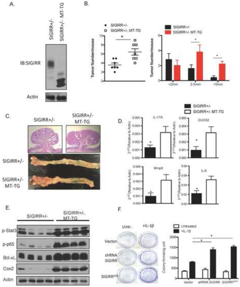
Figure 7. Loss of modification by complex glycan is sufficient to inactivate SIGIRR in tumorigenesis.
A. Colonic epithelial cell lysates from MT-SIGIRR, SIGIRR+/− mouse and SIGIRR+/− mouse was subjected to western blot analysis with anti-SIGIRR antibody. B. Mouse of indicated genotypes were subjected to AOM-DSS treatment. Tumor numbers and sizes were recorded and plotted. (N=7 for SIGIRR+/−, N=10 for MT-SIGIRR, SIGIRR+/−) C. Representative macroscopic view of colons from mice of indicated genotypes after the AOM-DSS treatment and H&E staining of tumors. D. Tumors from mice of indicated genotypes were subjected to real-time PCR analysis of indicated genes. E. Tumor lysates were subjected to western blot. Each lane represents one mouse. E. HT-29 cells were infected with lentivirus carrying an empty vector, shRNA targeting SIGIRR, or SIGIRR ΔE8 cDNA under a CMV promoter. The cells were cultured in the presence or absence of IL-1β for 5 days followed by crystal violet stain for formed colonies and colometric quantification of the colony formation. Error bar represents S.E.M * indicates p<0.05.

