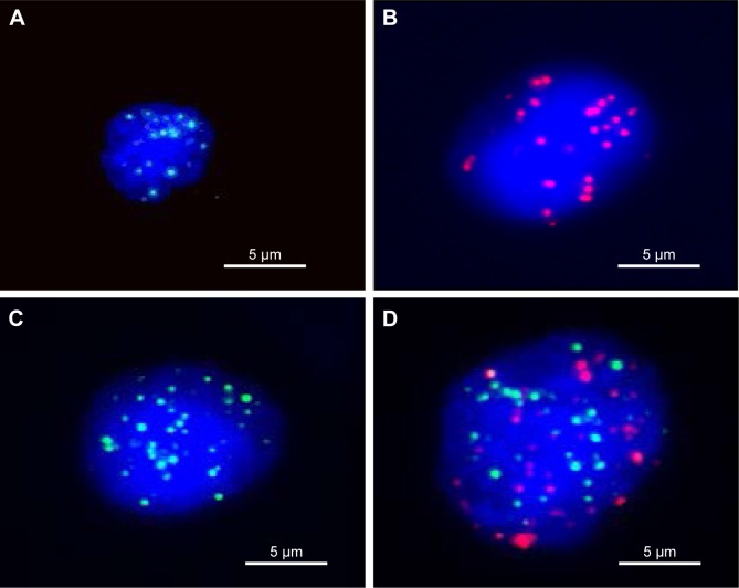
Figure 1.
Fluorescence microscopy images of three types of CTCs isolated from the peripheral blood osteosarcoma patients, based on RNA-ISH staining of epithelial (red dots) and mesenchymal (green dots) markers.
Notes: (A) White blood cells, (B) epithelial CTCs, (C) mesenchymal CTCs, (D) epithelial/mesenchymal CTCs. Scale bar, 5 μm.
Abbreviations: CTCs, circulating tumor cells; ISH, in situ hybridization.

