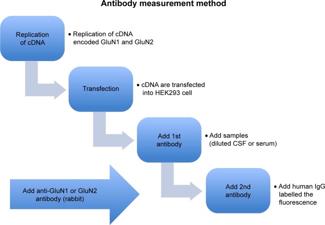
Figure 1.
Methods of anti-NMDAR antibodies detection.
Notes: The NMDAR subunit cDNAs (GluN1 alone or GluN1 together with GluN2B as an eqimolar mixture) were transfected with a lipofectamine reagent into human embryonic kidney (HEK) 293 cells. The cells were incubated with patient’s CSF, and then with FITC-conjugated anti-human IgG. To confirm the localization of NMDAR antibody binding sites, double staining was performed using both of the patients’ CSF and rabbit anti-GluN1 antibodies as the primary antibodies and a mixture of FITC-conjugated anti-human IgG and PE-conjugated anti-rabbit IgG as the second antibodies to confirm that the patient CSF really bind to GluN1.
Abbreviations: cDNAs, complementary DNA; CSF, cerebrospinal fluid; FITC, fluorescein isothiocyanate; NMDAR, N-methyl-D-aspartate receptor.

