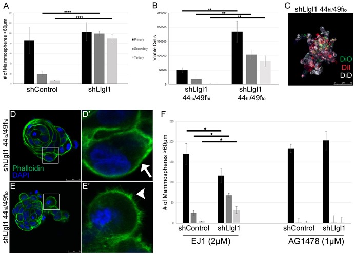
Figure 3. Loss of Llgl1 induces EGF-dependent mammosphere formation.

A.-F. Mammosphere assays were performed using cells generated as described in Figure 1 and Figure 2. A. Mammosphere formation with EGF treatment (20ng/mL) was quantified by counting mammospheres greater than 60μm (3 replicates per treatment group, each experiment was performed 3 times). B. Due to coalescing mammospheres in shLlgl1 CD44hi/CD49flo mammopshere formation was determined by counting viable cells at each time point (3 replicates per treatment group, each experiment was performed 3 times). C. MCF12A shLlgl1 CD44hi/CD49flo cells from primary mammospheres were labeled with the lipophilic tracer dyes Di-O, Di-I, or Di-D. D.-E. Mammospheres from shLlgl1 CD44lo/CD49fhi and CD44hi/CD49flo populations were incubated with Alexa Fluor 488 phalloidin (green) and DAPI (blue). The arrow indicates smooth cortical actin (D′) and the arrowhead indicates invasive cortical actin (E′). F. Mammospheres were treated with either 2μM EJ1 or 1μm AG1478. All primary passages are shown in black, secondary passages in dark gray, and tertiary passages in light gray. Error bars show ± standard deviation. *P < 0.05, **P < 0.01, ****P < 0.0001.
