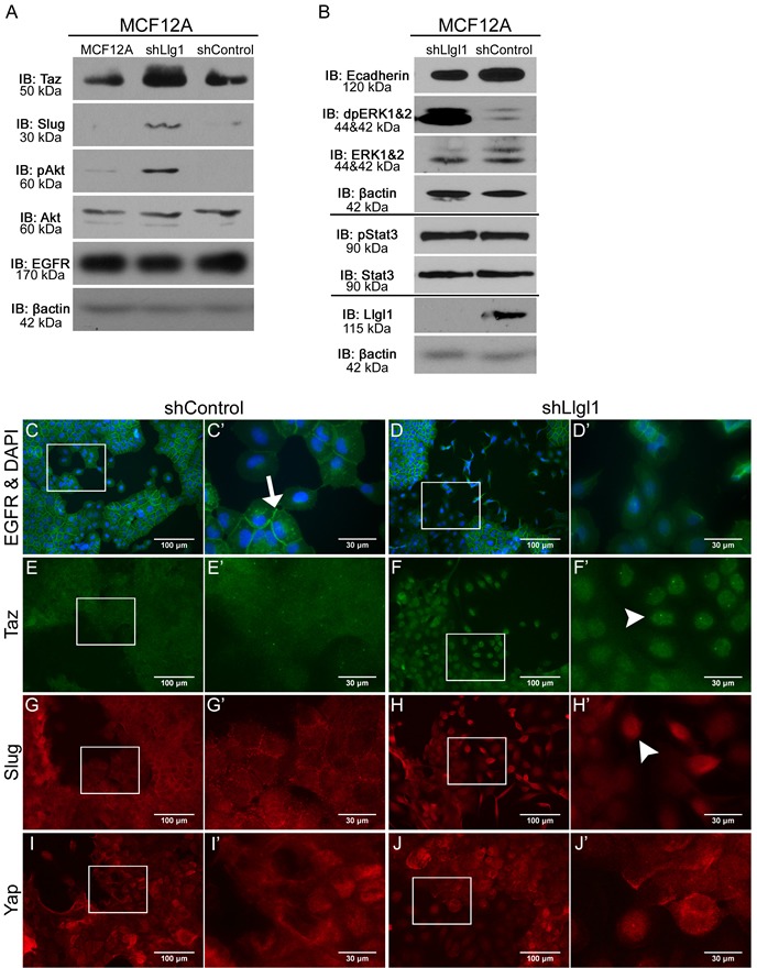
Figure 4. Llgl1 loss drives EGFR mislocalization and novel signal transduction.

A. and B. Protein lysates were collected from MCF12A parental, shControl, and shLlgl1 cells and analyzed by immunoblot using the antibodies: anti-TAZ, anti-Slug, anti-pAkt, anti-Akt, anti-EGFR, anti-E-cadherin, anti-dpERK1/2, anti-ERK1/2, anti-pStat3, anti-Stat3, anti-Llgl1, and anti-βactin. Immunoblots against anti-βactin are shown for each set of lysates. C.-J. MCF12A shControl and shLlgl1 cells were grown on plastic and (C-D) serum starved overnight or (E-J) in normal growth conditions then evaluated for localization of the indicated proteins. Cell were incubated with either anti-EGFR 1005, anti-TAZ, anti-SLUG, or anti-YAP antibodies and mounted (C-D) with DAPI or (E-J) without DAPI. Arrows indicate membrane localization and arrowheads indicate nuclear localization. C′, D′, E′, F′, G′, H′, I′, and J′ panels represent increased magnifications of C, D, E, F, G, H, I, and J panel insets.
