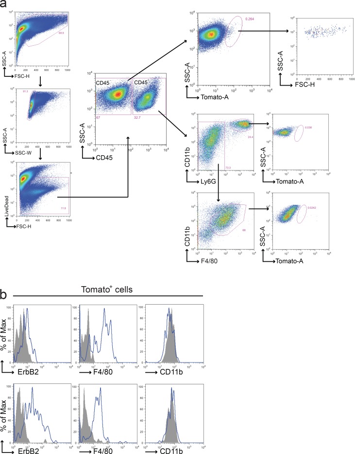
Figure 4. Fusion between cancer cells and macrophages in the BMT chimeric model.
a. Representative FACS analysis of tumors from Tomato/neu animals transplanted with CRE+ BM. Upon doublets and death cells exclusion, leukocytes were discriminated from tumor and stromal cells using anti-CD45 antibody. CD45− and CD45+ live populations were analyzed for the presence of Tomato+ cells. Tomato+ cells were found in CD45− population only and were characterized by a well-defined morphology (high FSC and SSC values) supporting the absence of debris in the gated region. Both macrophages and polymorphous nucleated cells presented in the CD45+ tumor population were negative for Tomato expression. Each gated region was defined using the appropriate FMO negative control. b. Representative flow cytometric analysis of ErbB2, F4/80 and CD11b on the Tomato+ sub-population derived from two distinct tumors. In agreement with the previous model, Tomato+ fused cells expressed ErbB2 and F4/80 on their surface but resulted negative for CD11b. Grey fill histograms represent isotype controls plus Fluorescence Minus One (FMO), while blue lines are ErbB2 or F4/80 or CD11b stained samples. Both macrophages and polymorphous nucleated cells presented in the CD45+ tumor population were negative for Tomato expression.

