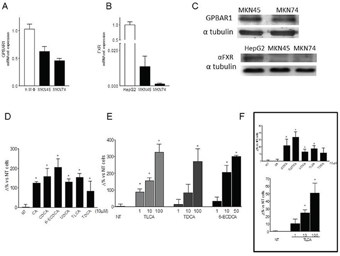
Figure 2.

A-B. Expression levels of GPBAR1(A) and FXR(B) mRNA were evalueted by ReaL-Time PCR analysis in MKN45 and MKN74 cell lines. Values are normalized to GAPDH and are expressed relative to those of positive controls (PBMC isolated from an healthy donor and HepG2 cellular line respectively), which were arbitrarily settled to 1. The relative mRNA expression is expressed as 2(−ΔΔCt). C. Western Blot analysis of GPBAR1 and FXR protein expression in gastric cancer cell lines. Data was normalized with α-Tubulin expression. Western blot shown is representative of two others showing the same pattern. D. Transwell migration assay. MKN45 were seeded, serum starved and then primed with 10 μM of cholic acid (CA), chenodeoxycholic acid (CDCA), ursodeoxycholic acid (UDCA), taurolithocholic acid (TLCA), taurodeoxycholic acid (TDCA) and 6-ECDCA. *P<0.05 versus not treated (NT) E. In another experimental setting, cells were treated with TLCA (1, 10 and 100μM), TDCA (1, 10 and 100μM), 6-ECDCA (1, 10 and 50μM) for 72 hours. Experiments were performed in triplicate. All three ligands increased, in a dose-response manner, migration activity of gastric cells in comparison with control cells. *P<0.05 versus not treated (NT). F. Cell adhesion to peritoneum. Experiments were conducted in triplicate. TLCA treatment significantly increased cellular adhesiveness of gastric tumor cells to murine parietal peritoneum in a dose dependent manner. *P<0.05 versus not treated (NT).
