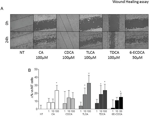
Figure 3. Wound healing assay.

A-B. Gastric cancer cells were serum starved and then primed with TLCA (1, 10 and 100 μM), TDCA (1, 10 and 100 μM), 6-ECDCA (1, 10 and 50 μM) for 72 hours. (A) A scratch wound healing assay is shown: MKN45 cell line left untreated or stimulated with 100 μM CA, CDCA, TLCA, TDCA and 50 μM 6E-CDCA. The images show cell migration after 72 hours of incubation with the indicated compound. After treatments with indicated compounds, cell monolayers were scraped in a straight line using a p200 pipette tip in order to create a “scratch”. A wound was generated and imaged at 0 an 24 hours with a phase-contrast. Images obtained from each samples at both time points were analyzed using Image J software and migration areas were expressed in pixels. All experiments were performed at least in triplicate. *P<0.05 versus not treated (NT).
