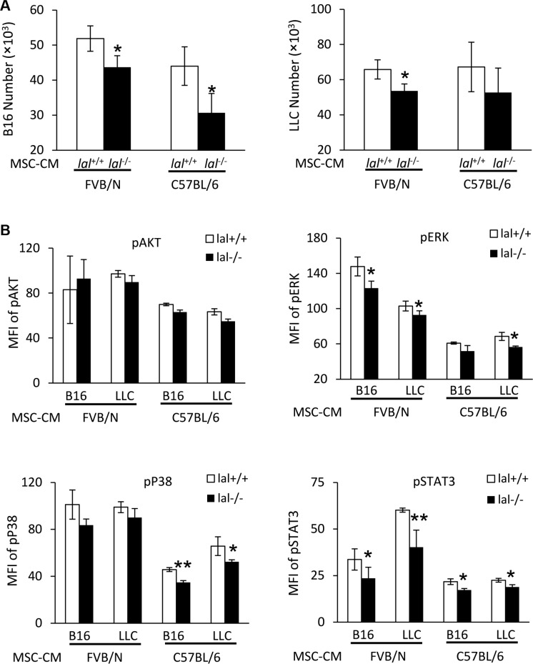
Figure 4. MSC-CM stimulates activation of intracellular signaling molecules.
(A) Stimulation of B16 melanoma cell proliferation by MSCs conditioned medium (CM). B16 melanoma cells (5 × 10³) or LLCs (1 × 10⁴) were seeded into 96-well plates, and then treated with CM from lal +/+ or lal −/− FVB/N or C57BL/6 MSCs. The cell number was counted at 72 h after CM treatment. (B) Activation of intracellular signaling molecules in B16 melanoma cells by MSCs CM. Two hours after CM treatment, B16 melanoma or LLC cells were harvested for flow cytometry analysis. lal −/− MSC-CM decreased phosphorylation of ERK, P38 and STAT3 in B16 melanoma and LLC cells. Statistical analysis of mean fluorescent intensity (MFI) by flow cytometry is shown. In the above experiments, data were expressed as mean ± SD; n = 3~4. *P < 0.05, **P < 0.01.

