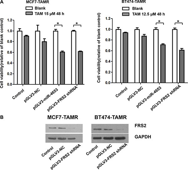
Figure 8. Overexpression of miR-4653-3p and knockdown of FRS2 enhanced the sensitivity to tamoxifen in MCF7-TAMR and BT474-TAMR cells.

MCF7-TAMR and BT474-TAMR cells were infected with lentivirus particles: pGLV3-miR-4653 which mediates pre-miR-4653 expression, pGLV3-FRS2 shRNA which interferes FRS2 expression, or the negative control (pGLV3-NC). (A) Cells were then exposed to TAM (12.5 or 15 μM) for 48 hours. Cell viability was detected by MTT assays. The bars represent the mean ± standard deviation of at least 3 independent experiments for each condition. * indicates significant inhibition of cell viability compared to controls (two-tailed t-test P < 0.05). (B) Cellular protein was isolated from TAM resistant cells followed by Western blot analysis with antibodies against FRS2 protein. GAPDH served as internal control. Control, indicates untransfected cells.
