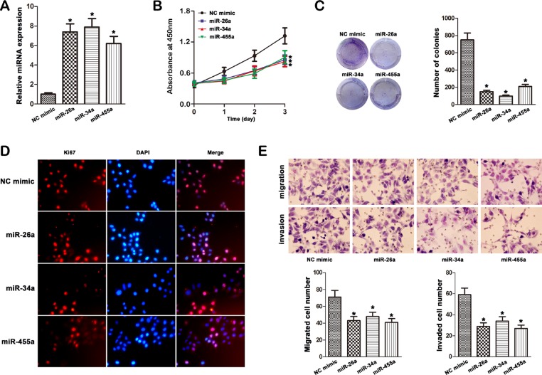
Figure 5. Effect of miR-26a, miR-34a and miR-455-3p mimic on cell progression in MHCC97H cells.
(A) The expression of miR-26a, miR-34a and miR-455-3p was studied by qRT–PCR in MHCC97H cells transfected with the mimics (*P < 0.05). (B) Transfection of miR-26a, miR-34a and miR-455-3p mimic in MHCC97H cells inhibited cellular viability as revealed by CCK-8 assay (*P < 0.05). (C) Overexpression of miR-26a, miR-34a and miR-455-3p inhibited the growth of MHCC97H cells with in focus formation assay. (D) Ki67 expression was detected by immunofluorescence staining in MHCC97H cells treated with miRNA mimic transfection. Red fluorescence: Ki67; DAPI staining for nuclear DNA. (E) Transwell cell migration and invasion assays were used to compare cell migration and invasion between miRNA mimic- and NC mimic-transfected cells. The data were mean ± S.D. of three separate transfections (*P < 0.05).

