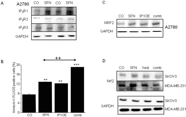
Figure 3. IP3Rs are involved in increased NRF2 expression due to SFN (20μM) treatment in A2780, SKOV3 and MDA-MB-231 cells.

SFN increases IP3R1, but not IP3R2 and IP3R3 protein amount A. in 104 and 105 A2780 cells, respectively. As a loading control, glyceraldehyde-3-phosphate dehydrogenase (GAPDH) was used. Further, we transfected cells with the IP3R1 (IP1OE) and determine NRF2 protein and the amount of apoptotic cells after a subsequent SFN treatment B, C. We observed significant increase in both, NRF2 protein (C) and apoptosis (B) compared to wild A2780 cells treated with SFN. Also, SFN-induced increase of NRF2 protein was observed in SKOV3 and MDA-MB-231 cells. This increase was prevented by parallel treatment with Xest and SFN D. Each column is displayed as mean ± S.E.M and represents an average of three independent cultivations, each performed in triplicates. Statistical significance ** compared to control represents p <0.01 and *** p <0.001. Statistical significance ++ compared to SFN treated cells represent p <0.01.
