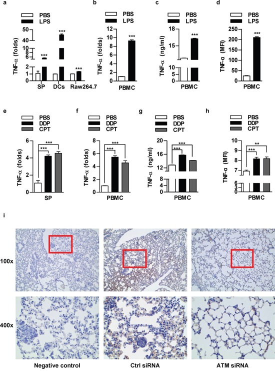
Figure 6. The components of lung cancer microenvironment could secret TNF-α in autocrine or paracrine manner upon LPS or chemotherapeutic drug stimulation.

Murine splenocytes, dendritic cells, Raw264.7 cells a, e. or human peripheral blood mononuclear cells (PBMC) b-d, f-h. was stimulated with LPS (a-d) or treated with DDP (1 μg/ml)/CPT (2 μg/ml) (e-h) and TNF-α expression was determined by RT-qPCR (a-b, e-f), ELISA (c, g) and flow cytometric analyse (d, h). The concentration of LPS for splenocytes, PBMC, Raw264.7, DCs was 100, 50, 50, 20 ng/ml respectively. Data are presented as the mean±SEM, n=3. **p<0.01; ***p<0.001, Student t test or one-way ANOVA with post Newman-Keuls test. One representative from three experiments is shown. SP: Splenocytes; DCs: dendritic cells. i. 8×10⁵ NCI-H520 cells conferred ATM silencing were transferred to BALB/c nude mice (5-6 weeks old) through tail vein (n=3 per group) and the lungs were performed TNF-α IHC staining.
