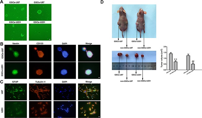
Figure 1. Isolation and identification of GSCs.

A. a and c: U87 and U251 glioma cells formed spheres in serum-free medium. b and d: single cells formed spheres again in a second-round sphere-forming assay. B. GSCs-U87 and GSCs-U251 stained for Nestin (green) and CD133 (red) by immunofluorescence analysis. C. GSC-U87 and GSC-U251 spheres were differentiated and then stained for GFAP (green) and beta-tubulin III (red) by immunofluorescence. D. Subcutaneously implanted GSCs-U87 or GSCs-U251 formed xenografts in nude mice. Data are presented as the mean ± SD (n=5, each group). **P<0.01 vs. GSC-U87 group; ##P<0.01 vs. GSC-U251 group.
