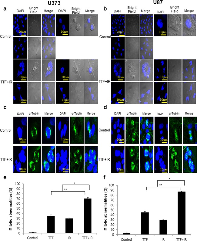
Figure 3. TTF+IR triggers multinucleation and mitotic abnormalities in glioblastoma cells.

Cells were exposed to 24 h of TTF, 5 Gy of γ-rays or 5 Gy of γ-rays followed by 24 h of TTF, indicated as the TTF, IR and TTF+IR treatments, respectively. a, b. Glioblastoma cells treated with TTF+IR. c, d. Immunofluorescence microscopy image of cells stained for α-tubulin (green) and DAPI. e, f. The histograms summarize the results of three independent experiments (with at least 100 cells counted in each experiment in each column). The values represent the means of three experiments ± SD; *p < 0.05, **p < 0.001. Cells were scored for the presence (abnormal) or absence (normal) of chromosome alignment and segregation defects. The image was acquired 24 h after the treatment was complete.
