
Figure 4. IL-6 activity is critical for lapatinib resistance.
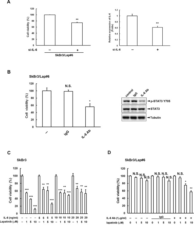
A. SkBr3/Lap#6 cells were transfected with si-control or si-IL6. Two days later, cells were re-seeded at a density of 2 × 10⁴ in a 6-cm dish for 2 weeks. Cell viability was detected and quantified by crystal violet staining. Gene silencing of IL6 was confirmed by ELISA. Statistical analysis was performed by Student's t test. **, p<0.01 as compared to control group. B. SkBr3/Lap#6 cells were cultured at a density of 1 × 10⁴ cells in 96-well plates with medium treated with either 10 μg/ml IgG or IL-6 antibody for 4 days. Cell viability was examined by MTT assay. Cell lysates treated with 10 μg/ml IL-6 antibody were also collected to examine the expression of phosphorylated STAT3 tyrosine 705 by western blot. Statistical analysis was performed by Student's t test. *, p<0.05 as compared to control group. N.S. represents non-significant. C. SkBr3 cells were pre-treated with different concentrations of recombinant IL-6 protein, followed by treatment with the indicated concentrations of lapatinib for 3 days. Cell viability was then examined by MTT assay. Statistical analysis was performed by Student's t test. **, p<0.01; ***, p<0.001 as compared to control group. D. SkBr3/Lap#6 cells were pre-treated with or without IL-6 antibody, followed by treatment with the indicated concentrations of lapatinib for 3 days. Cell viability was then examined by MTT assay. Statistical analysis was performed by Student's t test. *, p<0.05; **, p<0.01 as compared to each control group. N.S. represents non-significant.