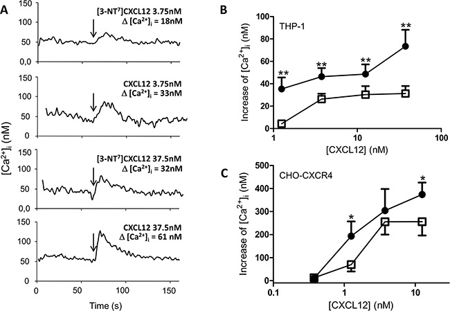
Figure 6. Calcium mobilization following CXCR4 activation with CXCL12 and [3-NT7]CXCL12.

A. Real-time changes of intracellular calcium levels (Δ[Ca2+]i) are shown in function of time. After 60 seconds, indicated by the arrows, 3.75nM or 37.5nM of CXCL12 or [3-NT7]CXCL12 are added to CHO-CXCR4 cells. The summarizing figures of these calcium mobilization experiments are shown in panel B and C. B, C. The increase of free intracellular calcium ions after stimulation of THP-1 cells (panel B; n= 6; ± SEM) or CHO-CXCR4 cells (panel C; n=7; ± SEM) with CXCL12 (•, filled circles) and [3-NT7]CXCL12 (□, open squares) was monitored (concentrations ranging from 0.375nM to 37.5nM).
