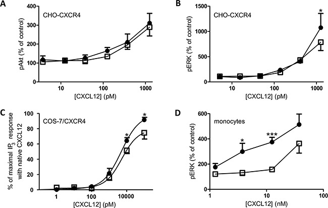
Figure 7. ERK, Akt and IP3 signal transduction pathways activated by CXCL12 and [3-NT7]CXCL12.

The accumulation of second messengers after stimulation of different cell types with CXCL12 (•, filled circles) and [3-NT7]CXCL12 (□, open squares) is shown. A, B. The average accumulation (n=8; mean ± SEM) of phosphorylated Akt and ERK1/2 after a two minute stimulation of CHO-CXCR4 cells with the CXCL12 forms (concentrations ranging from 3.75pM to 1.25nM) was calculated as a percentage compared to vehicle stimulated cells. C. IP3 generation (n=6; mean ± SEM) after stimulation of CXCR4-transfected COS-7 cells with both CXCL12 forms at concentrations ranging from 1pM to 100nM was measured. D. Fresh monocytes were stimulated with the CXCL12 forms (concentrations ranging from 1.25nM to 37.5nM). The average accumulation (n=9; mean ± SEM) of phosphorylated ERK1/2 after a two minute stimulation was calculated as a percentage compared to vehicle stimulated cells. All statistical analyses for differences between both CXCL12 forms were performed using the Mann-Whitney U test (* p < 0.05, ** p < 0.01, *** p<0.001).
