
Figure 5. Micelles-PTX combined with IMA pretreatment significantly inhibited the growth of tumors.
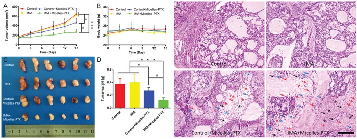
A. Tumor growth curve and B. mouse body weight curve throughout the experiment. C. Tumor xenografts images and D. tumor weights at the study end point. * P<0.05, *** P<0.001 compared with the control or IMA group, # P<0.01 compared with the Control+Micelles-PTX group. E. H & E staining of A549 xenograft slices from mice after various treatments. Black, blue and red arrows indicated karyopyknosis, karyorrhexis and karyolysis, respectively. The bar indicated 100 μm. Mouse models with size-matched A549 tumor xenografts were randomly assigned into four groups (n=6) and received oral treatment of IMA (50 mg/kg) or deionized water for two weeks followed by Micelles-PTX treatment. After Micelles-PTX treatment started, IMA or deionized water treatment was continued for another week. Micelles-PTX treatment was continued every third day for five times with the PTX dose of 5 mg/kg.