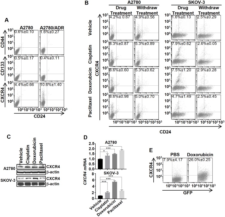
Fig 1. Chemotherapy transiently induced the CXCR4High cell state transition in human OVC.
(A) Representative FACS analysis showed that A2780/ADR displays a higher percentage of CXCR4High than A2780. Cells were stained with fluorescently labeled anti-CD44, anti-CD133, or anti-CXCR4, and also with anti-CD24. The numbers in the quadrants indicate the average percentage of CXCR4High. (B) Chemotherapy reversibly induced CXCR4High in A2780 and SKOV-3. A comparison of the changes in the cell population 72 h after treatment with suboptimal concentrations of cisplatin (1 μM), doxorubicin (10 nM), and paclitaxel (100 pM). At 72 h after withdrawing the drugs, the percentage of CXCR4High returned to the basal level. (C and D) The drug-induced CXCR4High was confirmed using western blot and real-time PCR analysis of the CXCR4 expression in the cell lysates. All the results represent means ± SD of three independent experiments (t-test, *p<0.05, ***p<0.001). (E) Doxorubicin induces tumoral CXCR4+-C. Mice (n = 3/group) were implanted with SKOV3/GFP-Luc (tagged with GFP and luciferase) and then treated with PBS (control) or doxorubicin (5mg/kg) via IV injection. After 72 h, the tumors were dissociated and analyzed for the percentage of CXCR4+-C by FACS analysis.

