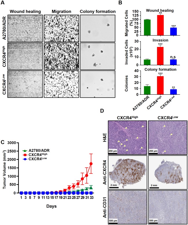
Fig 3. The freshly isolated CXCR4High showed enhanced invasion, migration, and tumor formation properties compared to the CXCR4Low.
(A) Microscopic images of wound healing, Boyden-chamber, and soft agar colony formation assays. (B) The results were also presented quantitatively to reflect the percentage and number of cells that invaded the wounds and migrated through the chambers, as well as the number of colonies formed. All results are presented as means ± SD of three independent experiments (t-test, **p<0.01 and ***p<0.001). (C) A comparison of the A2780/ADR, CXCR4High and CXCR4Low tumors’ growth rate in SCID mice (n = 5/group). The tumors were subcutaneously implanted on each side of the back of the animals. Data are presented as mean tumor volumes (mm3) ± SD versus time (two-way ANOVA, ***p<0.001). (D) Representative microscope images of the H&E staining and immunohistochemistry of the tumor sections show that CXCR4High tumor consist of higher expressions of CXCR4 and CD31 (blood vessel). Arrows indicate the vascular core areas.

