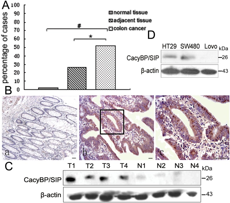
Fig 1. CacyBP/SIP expression in colon cancer and normal colon tissue.
(A), percentage of samples showing positive CacyBP/SIP immunoreactivity in normal colon samples, colon cancer tissue, and adjacent noncancerous colon tissue. Cells staining positive were defined as those showing cytoplasmic and/or nuclear staining. (B), representative photomicrographs of CacyBP/SIP staining in normal colon tissue (a, 100×), colon cancer tissue (b, 100×), and a close-up view of panel b showing diffuse cytoplasmic/nuclear staining (c, 400×). Bar = 50 μm. (C) and (D), Western blot analysis of CacyBP/SIP expression in colon cancer tissue and adjacent noncancerous tissue from four surgery patients, as well as the colon cancer cell lines HT29, SW480, and Lovo. All four colon cancer tissue samples (T1-T4) were positive for CacyBP/SIP expression, while the normal tissue samples (N1-N4) showed minimal expression. The colon cancer cell lines HT-29 and SW480 were positive for CacyBP/SIP expression, while the Lovo colon cancer cell line was negative. N, normal tissue; T, colon cancer tissue.

