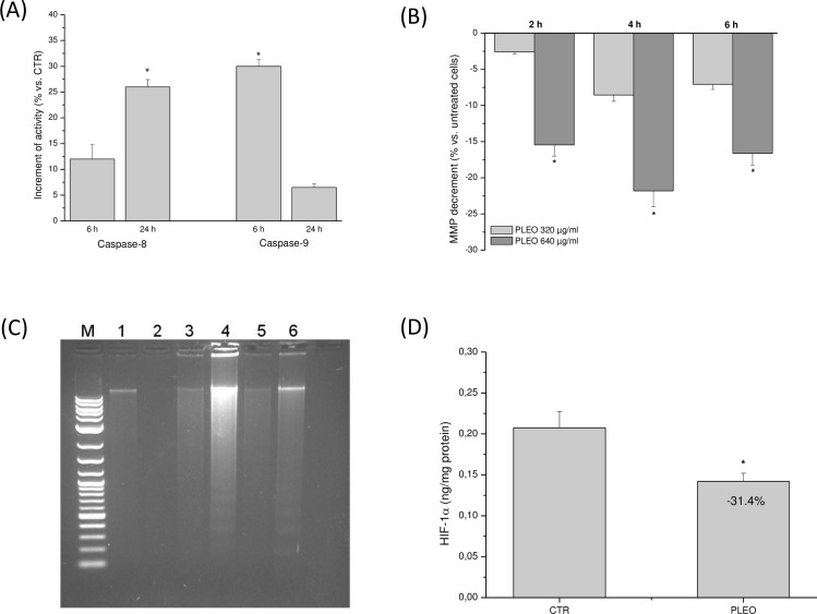
Fig 4.
(A) Caspase-8 and -9 activation after 6 and 24 h of PLEO administration (0.04% v/v, 320 μg/ml) to FTC-133 cells. (B) MMP reduction in FTC-133 cells incubated for 2, 4, and 6 h with PLEO (0.04–0.08% v/v, 320–640 μg/ml). (C) DNA fragmentation analysis in FTC-133 cells after 6, 24, and 48 h upon PL treatment (0.04% v/v, 320 μg/ml). M: molecular marker; 1–2: 6 h-CTR and PLEO-treated cells; 3–4: 24 h-CTR and PLEO-treated cells; 5–6: 48 h-CTR and PLEO-treated cells. (D) Reduction of HIF-1α levels after 24 h upon PLEO administration (0.04% v/v, 320 μg/ml) to FTC-133 cells. Data are expressed as mean ± SD (n = 3). *p<0.05 vs. untreated cells.

