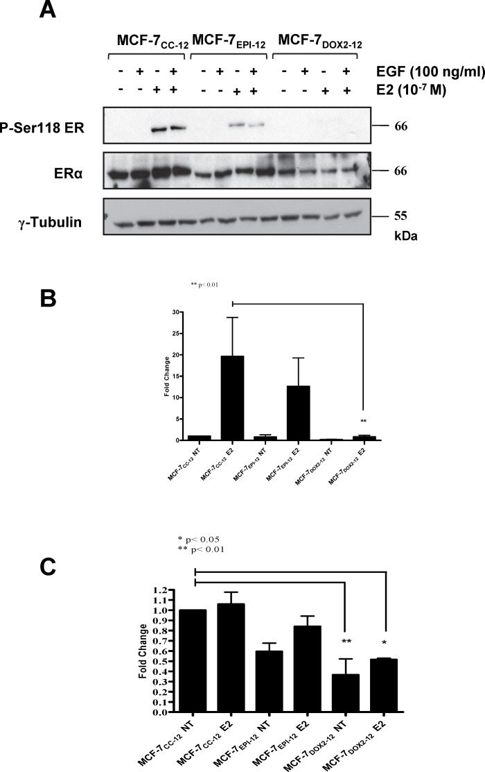
Fig 6. Differences in the expression of estrogen receptor alpha (ERα), phosphorylated estrogen receptor alpha at serine 118 (P-Ser118 ER), and γ tubulin in various cell lines, as measured in immunoblotting experiments with epitope- or phospho-specific antibodies.
(A) Immunoblots were conducted using extracts of cells with or without incubation in the presence of 100 nM E2, 100 mg/ml epidermal growth factor (EGF), or a combination of E2 and EGF. Primary antibodies purchased from Cell Signaling and used at 1:1000 dilution in 0.5% BSA overnight at 4°C. (B) Fold change in Ser-118 phosphorylated estrogen receptor relative to untreated MCF-7CC-12 cells, normalized to γ-tubulin expression (left) and to basal ERα expression (right). Data is expressed as the mean fold change observed in 3 independent experiments (± S.E.M., with the value of untreated MCF-7CC-12 cells set to 1.0. (C) Fold change in ERα levels, in a manner identical to that of P-Ser118 ER. The significance of differences between the test sample and that of untreated MCF-7CC-12 cells was assessed using an ANOVA test, followed by Bonferoni correction. * = p< 0.05, ** = p< 0.001

