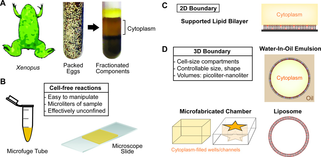
Figure 1. Xenopus Cell-free Egg Cytoplasmic Extracts and Strategies for 2D Confinement or 3D Encapsulation.
A. Eggs collected from the frog, Xenopus laevis, are fractionated to generate cell-free cytoplasm
B. The cytoplasmic extract is capable of carrying out complex cell biological processes in vitro in the absence of cell boundaries. Microliter volumes of cytoplasm are often activated and imaged in test tubes and on microscope slides.
C. Configuration for confinement of cellular reactions to a supported lipid bilayer surrounded by cytoplasm. The process of interest is initiated at or signals to a two-dimensional membrane whose composition is controllable.
D. Encapsulation of cytoplasmic extract reactions within cell-like boundaries. These stiffness of these boundaries varies: PDMS wells (rigid), water-in-oil emulsion (intermediate, lipid monolayer), or liposomes (soft, lipid bilayers). Importantly, compartment dimensions can be specified to encapsulate cell-size volumes of material; from picoliters to nanoliters

