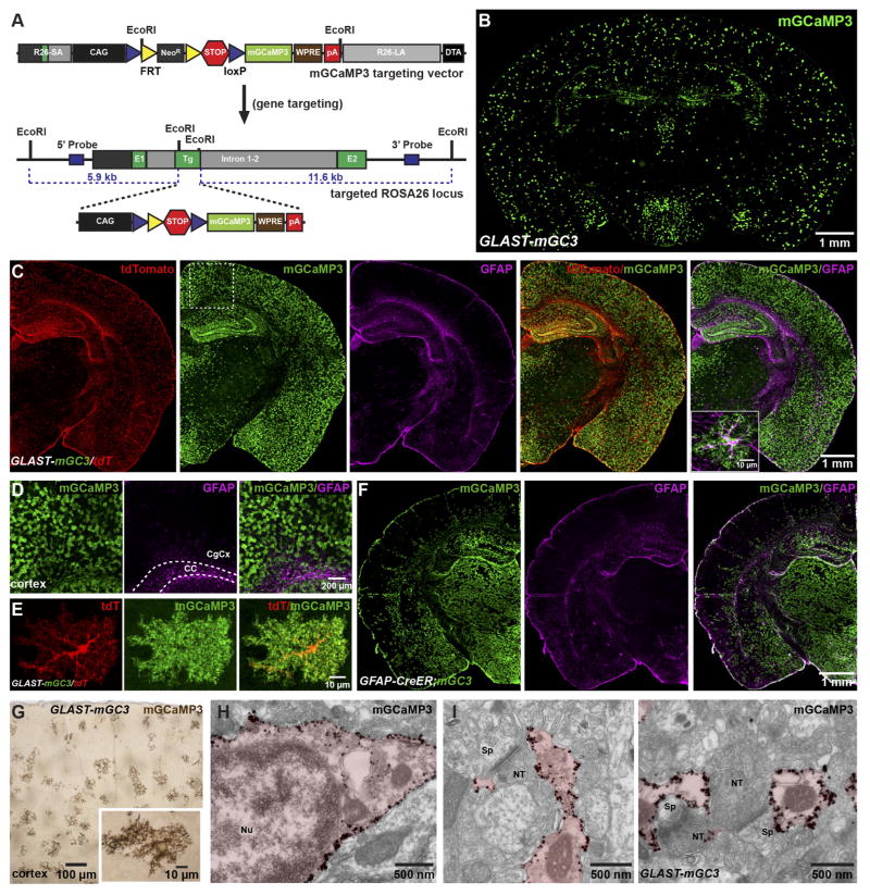
Figure 1. Conditional expression of mGCaMP3 in astrocytes.
(A) Gene trap strategy used to insert conditional mGCaMP3 transgene into murine Rosa26 gene. (Top) mGCaMP3 targeting vector which harbors 5′ short (R26-SA) and 3′ long (R26-LA) homology arms from Rosa26, the CMV enhancer chicken β-actin hybrid (CAG) promoter (black box), loxP (blue triangles) flanked neomycin resistance cassette (NeoR, gray box) flanked by two FRT sites (yellow triangles) and 3x SV40-polyA sequence (red hexagon), the mGCaMP3 cDNA (light green box), woodchuck post-transcriptional response element (WPRE, brown box), bovine growth hormone poly A sequence (red box) and negative selection cassette with diphtheria toxin fragment A (DTA, black box). (Bottom) Rosa26 allele with conditional mGCaMP3 cassette after homologous recombination. NeoR cassette was removed by in vivo site-specific recombination.
(B) Coronal section of brain from a GLAST-CreER;Rosa26-lsl-mGCaMP3 (GLAST-mGC3) mouse immunostained for mGCaMP3.
(C) Coronal hemi-section of brain from a GLAST-CreER;Rosa26-lsl-mGCaMP3;Rosa26-lsl-tdTomato (GLAST-mGC3/tdT) mouse immunostained for tdTomato, mGCaMP3 and GFAP. Inset shows one cortical astrocyte at higher magnification co-expressing mGCaMP3 and GFAP.
(D) High magnification images from boxed area in (C). CC: corpus callosum, CgCx: cingulate cortex.
(E) Images showing one cortical astrocyte from a GLAST-mGC3/tdT mouse (maximum intensity projected confocal z-stack) immunostained for mGCaMP3 and tdT.
(F) Coronal hemi-section of brain from a GFAP-CreER;Rosa26-lsl-mGCaMP3 (GFAP-CreER;mGC3) mouse immunostained for mGCaMP3 and GFAP.
(G) Coronal section of brain from a GLAST-mGC3 mouse showing silver intensified immunogold labeling of mGCaMP3. Inset shows one astrocyte at higher magnification.
(H, I) High magnification images of silver intensified immunogold labeling of mGCaMP3 in an astrocyte soma (H) and peri-synaptic processes (I) from GLAST-mGC3 mice. NT: nerve terminal, Nu: nucleus, Sp: spine.

