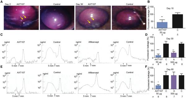
Fig. 4. Intraocular injection of AXT107 reduces VEGF-induced leakage for at least 2 months in rabbit eyes.
(A) Fundus photography 2 days after intraocular injection of 50 μg of AXT107 in Dutch Belted rabbits. The vitreous was clear with no evidence of inflammation, and a gel-like depot was seen in the inferior vitreous at the site of injection (left panel, arrows) and was still seen in the same location 30 days after injection (third panel, arrows). (B) VEGF (10 μg) was injected in both eyes3 days after injection of 50 μg of AXT107 in one eye, and after 7 days, VFP showed fluorescein leakage reduced by 60% in AXT107-injected eyes (**P = 0.007 by unpaired t test). (C) In another group of rabbits, 10 μg of VEGF was injected in both eyes 23 days after injection of 50 μg of AXT107 or 500 μg of aflibercept in one eye, and at 30 days, VFP scans showed low levels of sodium fluorescein in the mid-vitreous between 5 and 7 mm in front of the retina in AXT107- or aflibercept-injected eyes compared with fellow control eyes. (D) Quantification of VFP images in (C) showed that leakage was reduced by 86% in AXT107-injected eyes and 69% in aflibercept-injected eyes (**P < 0.01, ***P < 0.001 by unpaired t test from corresponding controls). (E) The rabbits from (D) were rescanned without injection of sodium fluorescein on day 43, and vitreous fluorescence was back to baseline levels. On day 53, 10 μg of VEGF was injected into each eye, and on day 60, VFP scans showed low levels of fluorescein in the mid-vitreous of AXT107-injected eyes and higher levels in control and aflibercept-injected eyes. (F) Quantification of VFP images in (E) showed that leakage was reduced by 70% in AXT107-injected eyes compared to corresponding controls (*P = 0.035 by unpaired t test) but not in aflibercept-injected eyes (A). Bars represent mean (± SEM).

