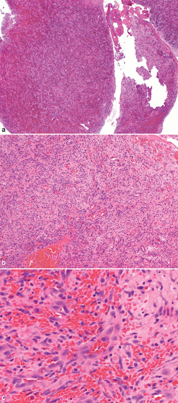
Fig. 3.

(a) Low-power histopathologic view displaying lesion with spindled cells and abundant pale cytoplasm. (b) Medium-power histopathologic view with better representation of spindled cells, abundant pale cytoplasm, and numerous erythrocytes. (c) High-power histopathologic view outlining vascular channels with generally smooth endothelial lining, scattered erythrocytes, and spindled cells.
