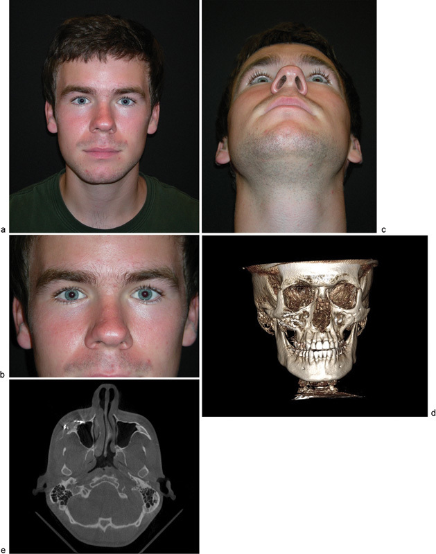
Fig. 5.

(a) 18-month postoperative view of the patient. (b) 18-month postoperative close-up of the bilateral orbitozygomatic region. (c) 18-month postoperative inferior view indicating acceptable uniformity of zygoma projection. (d) 18-month postoperative computed tomographic (CT) scan with 3D reconstruction. (e) 18-month postoperative CT axial scan indicating no evidence of recurrence of lesion.
