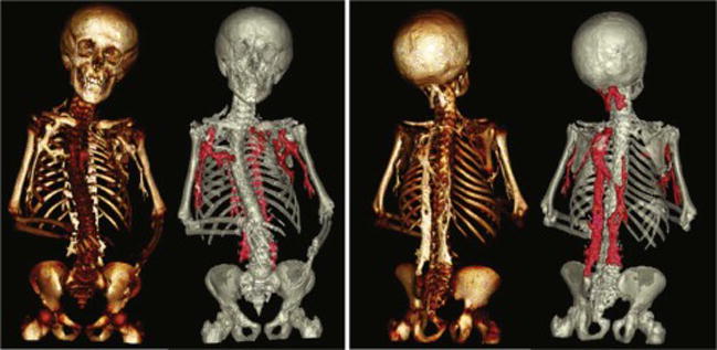
Figure 7.

Anterior and posterior 3D reconstructions of CT images obtained from an FOP patient with HO lesions highlighted in red. 3D reconstructions of CT images can aid in identifying lower grade lesions and in determining how lesions connect to and impact skeletal structure on a systemic level.
