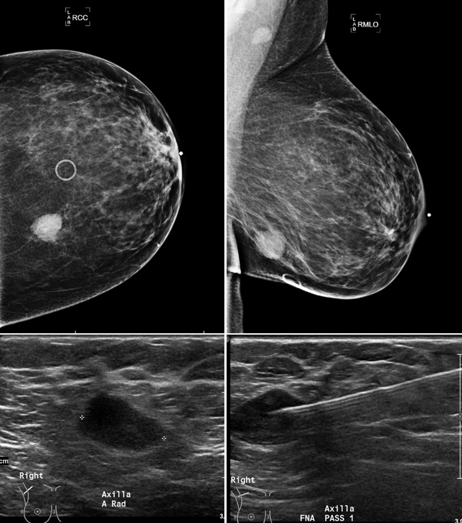
Figure 3.
48 year old female with a lower inner quadrant invasive ductal carcinoma (T2 tumor–2.1 × 1.6 cm mass, grade-3) (top row). Right axilla (RMLO view) demonstrates an oval heterogeneous anechoic lymph node (left bottom), without fatty hila, cortical thickening and abnormal vascularity (image not shown). FNA (needle in appropriate position, bottom right) was positive.

