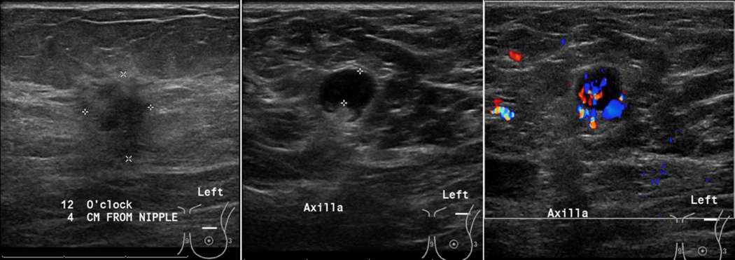
Figure 4.
75 year old female, with a left breast primary tumor (T1 – 1.4 × 1 cm at 12:00 clock) that was pathology-verified as invasive ductal carcinoma. Evaluation of the axilla demonstrated a round LN, with eccentric displacement of central fatty hila, focal cortical thickness of 5mm and non-hilar blood flow. FNA was positive for malignant cells of ductal origin.

