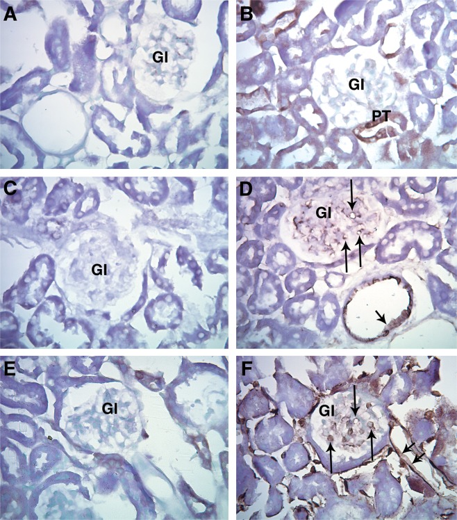
Figure 2.

Representative light micrographs of paraffin embedded renal cortex immunohistochemically stained for MT (A, B, E, F) and PECAM‐1 (C, D.). A. FVB: Negative control, primary antibody omitted. B. FVB: Endogenous MT staining was not observed in the vasculature, but was occasionally observed in proximal tubules (PT). C. FVB: Negative control, primary antibody omitted. D. FVB Staining was observed in the cytoplasm of EC in the capillaries of the glomerulus (arrow) and in renal cortical arterioles (arrowhead). E. JTMT: Negative control, primary antibody omitted. F. JTMT: Cytoplasm of glomerular capillaries (arrow), peritubular capillaries (double arrowheads), and and other cortical microvascular structures displayed intense MT stain. Gl; glomerulus.
