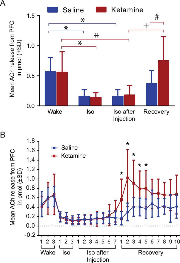
Figure 7. Mean acetylcholine (ACh) release in the prefrontal cortex during saline and ketamine injection.
A. No difference was seen between the two treatment groups during the wake phase. ACh release in the prefrontal cortex was significantly decreased by isoflurane administration (*). Injection of ketamine during isoflurane anesthesia did not result in any change of ACh levels compared to saline. However, after isoflurane was discontinued, ACh significantly increased (+) in ketamine treated animals (n=8) during recovery phase while animals in the saline group showed no significant difference. Compared to the saline group (n=9), animals injected with ketamine showed a significant increase in ACh release during the recovery phase (#). Data are shown as mean ACh release in pmol from prefrontal cortex (+SD). B. ACh release timeline in the prefrontal cortex. No difference was seen between saline and ketamine injection during the wake, isoflurane, and isoflurane after injection phase. However, ketamine treated animals showed a significant increase in ACh release within the prefrontal cortex for the first 62.5 min during the recovery phase after discontinuation of isoflurane. Data are shown as mean ACh release from the prefrontal cortex (± SD). Iso: Isoflurane; PFC: prefrontal cortex. Blue line: saline treatment, red line: ketamine treatment; statistical significance (*, +, #) defined as p <0.05.

