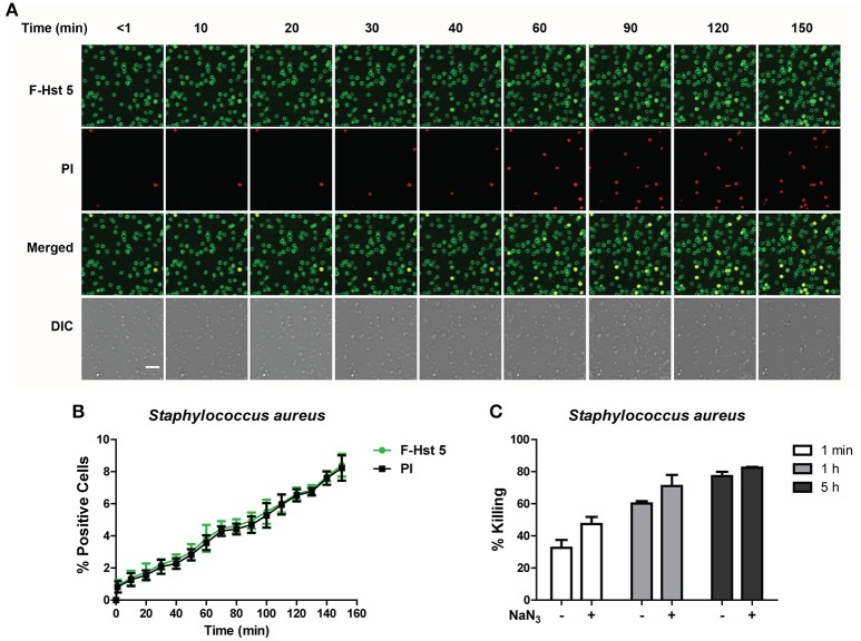
Figure 6.
The activity of Hst 5 against S. aureus is mediated by energy-independent mechanisms. (A) S. aureus cells were exposed to F-Hst 5 (30 μM) and PI (2 μg/mL). F-Hst 5 (green) and PI (red) uptake were measured in parallel by time-lapse confocal microscopy. Images were recorded every 10 min and selected images of indicated time points were shown. (Scale bar: 5 μm). (B) Quantitative analysis of F-Hst 5 uptake (green line) and PI uptake. Error bars represent the standard errors from four different fields of image. (C) Cells pretreated with 10 mM NaN3 at 37°C for 3 h did not show significant difference in susceptibility to Hst 5.

