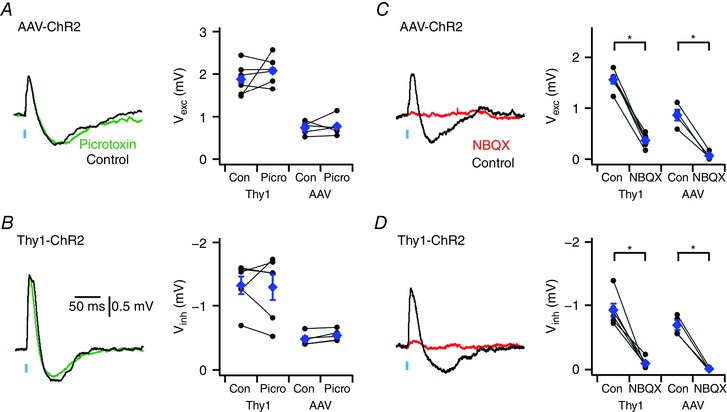
Figure 4. Depolarizing and hyperpolarizing components of synaptic responses are mediated by glutamate receptors.

A and B, examples (left) of synaptic responses to optical activation of neocortical inputs (A) and inputs in Thy1‐ChR2‐YFP mice (B) before (black traces) and during block of GABAA receptors with picrotoxin (green traces). Population data (right) indicate that picrotoxin does not modify excitatory (P = 0.89 and P = 0.32, for AAV (n = 5, N = 5) and Thy1‐ChR2‐YFP (n = 6, N = 6) experiments, respectively, paired t test) or inhibitory components (P = 0.57 and P = 0.86, paired t test). C and D, examples (left) of synaptic responses to optical stimulation of motor cortex neurones expressing AAV‐ChR2‐Venus (C) and of inputs in Thy1‐ChR2 mice (D) before and during block of GluA receptors with NBQX. Population data (right) indicate that NBQX abolishes excitatory (P = 0.017, n = 4, N = 4 and P = 9.5 × 10⁻⁶, n = 6, N = 6, paired t test) and inhibitory components (P = 0.033, n = 4, N = 4 and P = 8.6 × 10⁻⁴, n = 6, N = 6, paired t test). Data are presented as means ± SEM. * P < 0.05 control vs. NBQX (paired t test).
