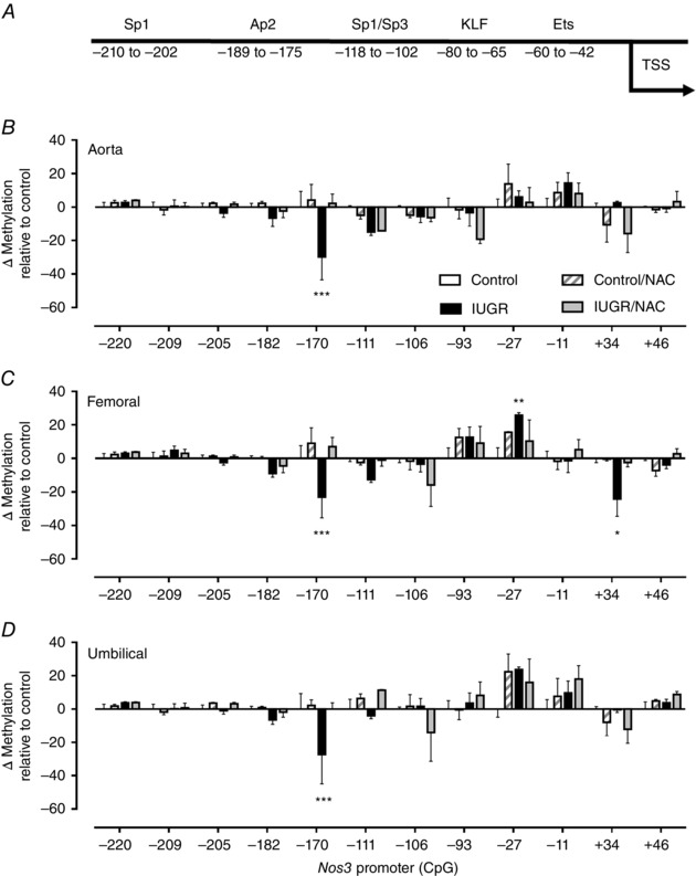
Figure 10. Level of DNA methylation in the Nos3 promoter of guinea pig fetal artery endothelial cells.

A, schematic representation of guinea pig Nos3 promoter and cognate binding sites for transcription factors predicted with MatInspector. B–D, change in DNA methylation levels relative to control in CpGs present in the Nos3 promoter in primary cultures of fetal ECs from aorta (B), femoral (C) and umbilical (D) arteries in untreated control (open bars) and IUGR (solid bars), as well as treated control (dashed bars) and IUGR (grey bars) guinea pigs. Values expressed as mean ± SEM, * P < 0.05, ** P < 0.01, *** P < 0.001 vs untreated control, two‐way ANOVA, Newman–Keuls multiple comparison test.
