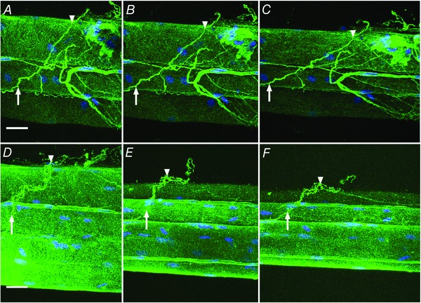
Figure 2. Collagen cables are composed of type I collagen and realign with increased strain.

Bundles of wild‐type muscle fibres were observed by confocal microscopy in a custom stretching chamber (n = 4). Bundle preparations (two examples shown in A–C and D–F) were immunolabelled for type I collagen (green) and nuclei (blue). At slack length (0% strain; A and D) intensely stained type I collagen positive structures that traverse multiple fibres are defined as collagen cables. Two points on a cable are identified in each preparation (arrow and arrowhead) and these points are again identified at 20% strain (B and E) and 40% strain (C and F) showing reorganization of cables with strain. Scale bar = 25 μm. [Colour figure can be viewed at wileyonlinelibrary.com]
