
Figure 1. Course of study.
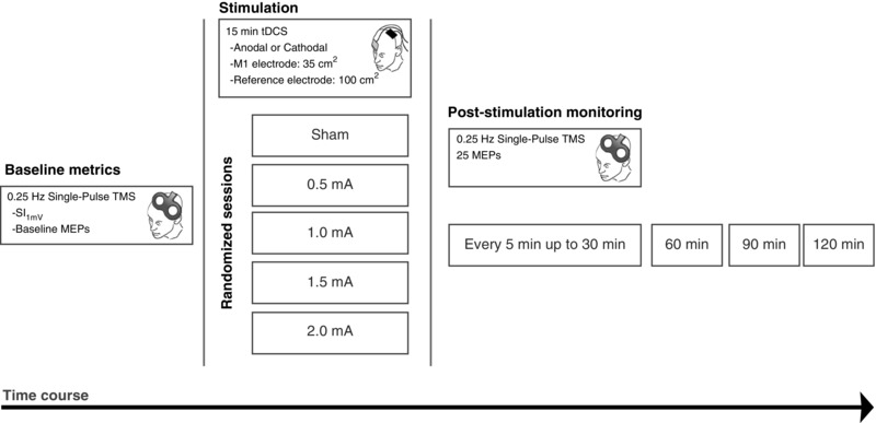
Participants were randomly divided into two groups for tDCS polarity (Anodal: n = 20; Cathodal: n = 18). Each participant took part in five randomized sessions during which either sham, 0.5, 1.0, 1.5 or 2.0 mA stimulation with the respective polarity was applied. Prior to receiving stimulation, baseline MEP amplitude and SI1mV was measured over the determined motor cortical ‘hotspot’ which produced the largest MEP from the right ADM muscle. Next, DC stimulation for 15 min was delivered, and MEP measurements were taken again from the hotspot immediately after stimulation, as well as every 5 min up to 30 min, and then every 30 min up to 2 h after stimulation.