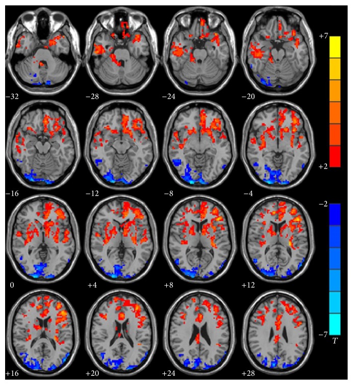
Figure 1.
Group analysis of ALFF between infants with CSSHL and healthy controls. Compared with the control group, decreased (red) ALFF was observed in left Heschl's gyri (BA41) (p = 0.0059, ES = 0.94), left superior temporal gyrus (BA22) (p = 0.0003, ES = 1.27), left inferior frontal gyrus (BA44) (p = 0.0004, ES = 1.27), left inferior frontal gyrus (BA45) (p = 0.0000, ES = 2.18), left inferior prefrontal gyrus (BA47) (p = 0.0001, ES = 1.40), and left dorsolateral prefrontal cortex (BA46) (p = 0.0000, ES = 1.68). Increased (blue) ALFF was observed in right occipital lobe and right angular gyrus, which included BA18 (p = 0.0022, ES = −1.07), BA19 (p = 0.0027, ES = −1.04), BA17 (p = 0.0000, ES = −1.46), and BA39 (p = 0.0020, ES = −1.08). AlphaSim corrected (p < 0.05, cluster size > 228 voxels). The significance level of activity was indicated by the color bar (T), increasing as red proceeding to yellow decreasing as blue to cyan.

