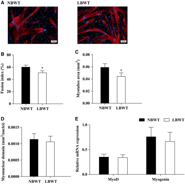
Figure 3.

Myogenic differentiation of satellite cells from LBWT and NBWT neonatal pigs. (A) Immunolabelling in satellite cell‐derived myotubes at day 3 of differentiation (Scale bar = 100 μm). (B) Fusion index expressed as the percentage of total nuclei that are located in myotubes stained in red. (C) Myotubes size expressed as the area (mm2) of myotubes that are myosin immunopositive (stained in red). (D) Myonuclei domain calculated as the area of myotubes (stained in red) divided by the number of DAPI‐stained nuclei inside. (E) mRNA expression of MyoD and Myogenin in myotubes of LBWT and NBWT neonatal pigs. Results are means ± SE. n = 8. *P ≤ 0.05 versus NBWT pigs. LBWT, low birth weight; NBWT, normal birth weight.
