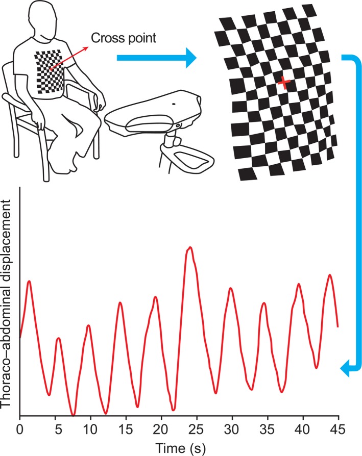
Figure 1.

Structured light plethysmography projects a grid of light onto the thoraco–abdominal (TA) wall of a participant. The cross point of the grid is centered at the base of the xiphisternum. Changes in the grid pattern are recorded using two cameras (located in the scanning head) and then translated into a virtual surface corresponding to the shape of TA wall of the subject. Average axial displacement of the virtual grid provides a one‐dimensional movement–over‐time trace from which tidal breathing parameters can be calculated. Modified from Elshafie et al. 2016. s, seconds.
