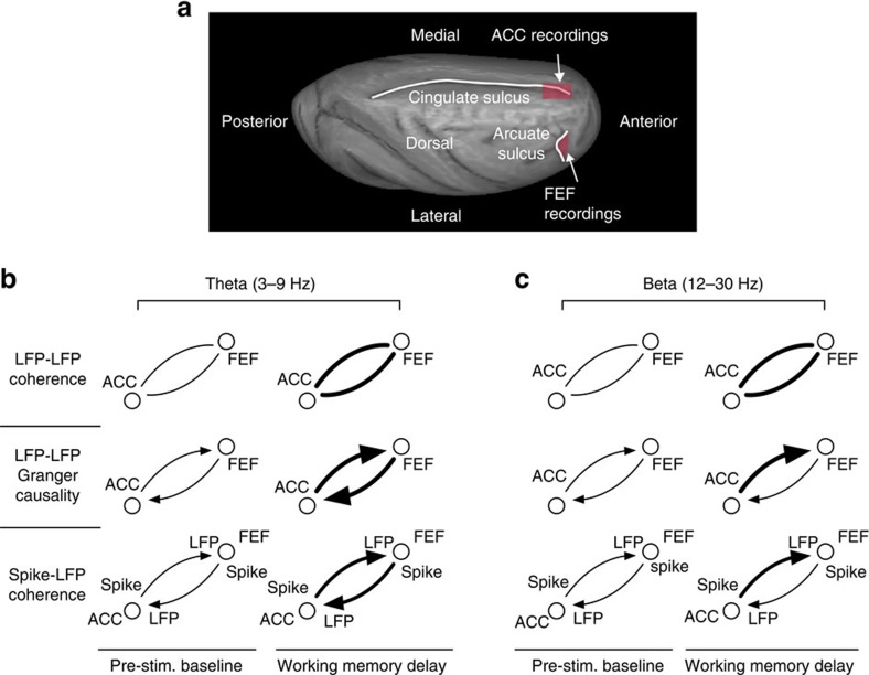
Figure 8. Illustration of recorded brain area locations and summary of main interareal ACC-FEF modulations observed in this study.
(a) ACC and FEF recording locations (in red shading) shown on a rendering of a semi-inflated macaque brain. (b,c) Illustration of main interareal effects in the theta (b) and beta (c) band with the thickness of connections indicating the strength or prevalence of the effects. LFP-LFP coherence (top row) was modulated during the delay in >75% of LFP-LFP pairs in both frequencies (with increased coherence in the largest majority). Granger causality (middle row) increased during the delay for both ACC to FEF, and FEF to ACC directions (more pronounced in theta band), but the ACC to FEF Granger-causal flow was stronger than FEF to ACC Granger-causal flow at both theta and beta frequencies. Spike-LFP coherence (bottom row) increased for both directions during the delay in the theta band, but was different between delay and baseline merely in one beta frequency bin (at 22 Hz) for ACC spike to FEF LFP sites for contraversive saccades. Reduced interareal modulation prior to error commission was evident in both frequencies across different measures and is described in the text.

