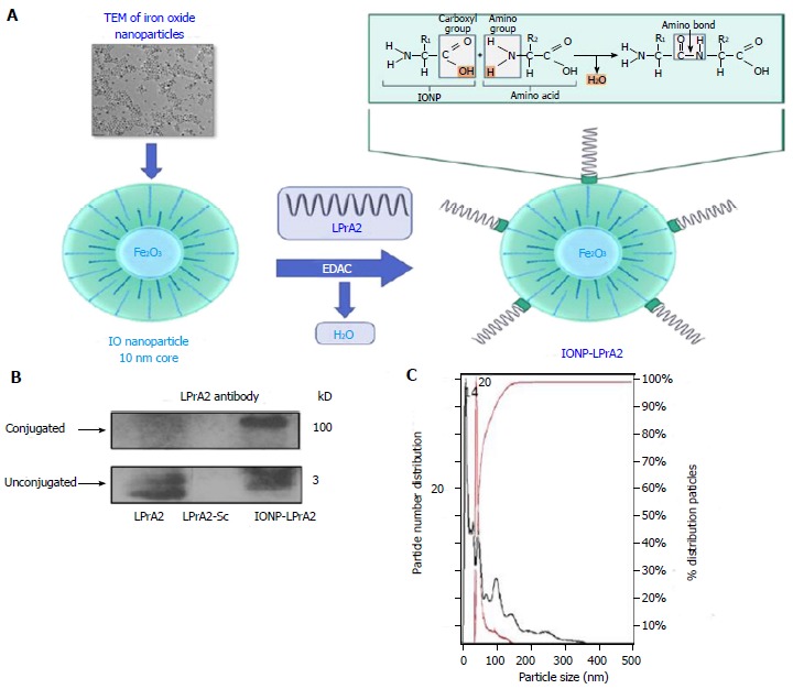
Figure 1.

Generation and characterization of IONP-LPrA2. A: Conjugation of IONP-LPrA2. LPrA2 was conjugated to IONPs via 1-Ethyl-3-(3-dimethylaminopropyl)carbodiimide (EDAC), which activates the carboxyl group on the IONP surface allowing it to form a covalent bond with the amino group of LPrA2 (displayed by TEM, transmission electron microscopy, Ocean Nanotech); B: Western blot confirmation of IONP-LPrA2 conjugation. Conjugated IONP-LPrA2 (100 kD) was detected by Western blot with an LPrA2 antibody, purified from antigen injected rabbit bleeds. Unconjugated LPrA2 (3 kD) and the scrambled peptide LPrA2-Sc (3 kD) served as positive and negative controls, respectively; C: NanoSight analysis of unconjugated and conjugated IONPs. The particle size of unconjugated IONP (14 nm) shown in black and the conjugated IONP-LPrA2 (20 nm) shown in red were determined by nanoparticle tracking analysis. The hyperbolic curve shows that the particles are 100% homogeneous.
