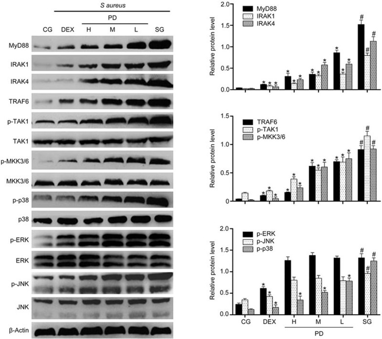
Figure 8.
The effects of PD on the TLR2-mediated activation of the MAPK signaling pathway in mMECs. The expression of MyD88, IRAK1, IRAK4, TRAF6, TAK1, MKK3/6 and MAPK protein in mMECs was determined using Western blotting. β-Actin was used as a control. CG is the control group; SG is the S aureus group; DEX is the dexamethasone group; and H, M and L are the PD administration groups, with doses of 45, 30 and 15 mg/kg per animal, respectively, and 100, 50 and 25 μg/mL in the cells, respectively. The data are presented as the mean±SEM. *P<0.05 vs SG. #P<0.05 vs CG.

