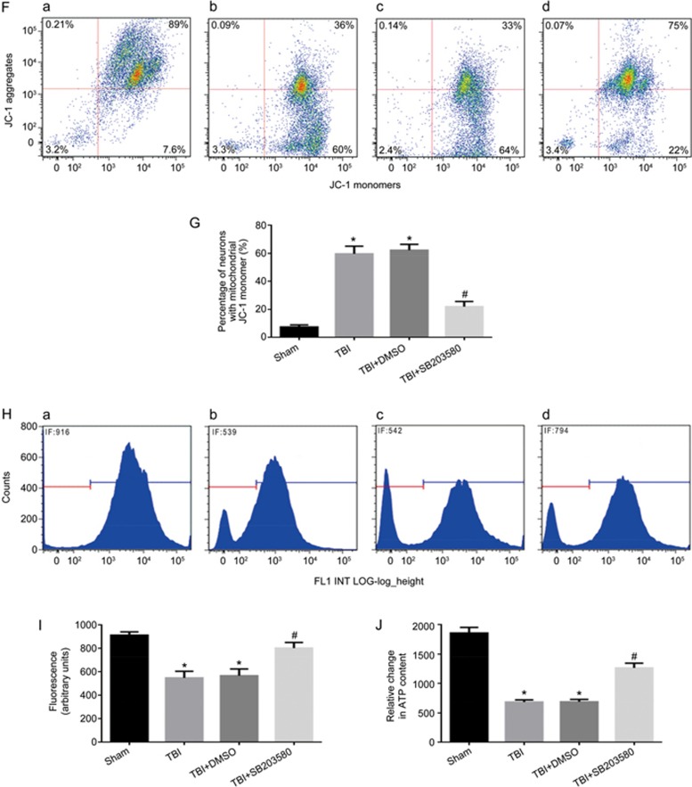
Figure 6F-6J.
Inhibition of p-p38 can alleviate mitochondrial damage and neuronal apoptosis. Rats were injected ip with the p38 inhibitor SB203580 (200 μg/kg) 30 min before TBI induced by LFP. After 12 h, rats were sacrificed. (F) The loss of ΔΨm was measured by JC-1 and analyzed by flow cytometry. (a–d) represent the sham group, TBI group, TBI+DMSO group and TBI+SB203580 group, respectively. (J) Quantification of mitochondrial depolarization expressed as JC-1 monomer (green fluorescence) of all treatment groups post-TBI. (H) mPTP opening was measured by staining with calcein-AM and CoCl2 and analyzed by flow cytometry for green fluorescence. (a–d) represent the sham group, TBI group, TBI+DMSO group and TBI+ SB203580 group, respectively. (I) Quantification of mitochondrial green fluorescence intensity post-TBI of all treatment groups. (G) Quantification of the change in neuronal ATP levels following TBI of all treatment groups. There was no difference between the TBI and the TBI+DMSO groups. P-38 inhibitor decreased the low ΔΨm (F, G) and the opening of mPTP (H, I), and increased neuronal ATP levels (J). n=6/group. Mean±SD. *P<0.05 vs sham group. #P<0.05 vs TBI group.

